
A picosecond ultrasonic pulse typically has a spatial width of less than 0.1 μm, making it smaller than the wavelength of visible light. Such pulses reflect light, acting like a semitransparent mirror. This allows us to follow the ultrasonic pulse by use of a reflected light beam (a probe beam), which undergoes oscillations.
This frequency of these oscillations can be used to determine the sound speed. Our previous research used this method to image animal cells. See 3D animal-cell imaging with picosecond ultrasonics.
In a recent study we have made two improvements to the detection technique in such experiments.
In the first improvement we use a high numerical aperture objective lens to change the angle of the light reaching the sample, thus allowing a range of ultrasonic frequencies to be probed. For more details see 'Time-domain Brillouin imaging of sound velocity and refractive index using automated angle scanning,' M. Tomoda, A. Kubota, O. Matsuda, Y. Sugawara, and O. B. Wright, Photoacoustics. 31, 100486 (2023).
Schematic of the angle scanning method (the left-hand figure) and graph of reflectance change vs probe light position on the objective lens (related to the angle) and delay time (the right-hand figure)
The second improvement involves a setup that allows sound velocity changes to be monitored without the need for a separate measurement of refractive index. 'Sound velocity mapping from GHz Brillouin oscillations in transparent materials by optical incidence from the side of the sample,' M. Tomoda, A. Toda, O. Matsuda, V. E. Gusev, and O. B. Wright, Photoacoustics. 30, 100459 (2023).
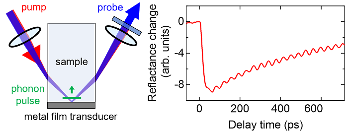
Schematic of the our method involving optical incidence from the side of the sample (left) and the measured reflectance change (right).