See also the layperson's introduction: Jumping frogs, squeaking bats and tapping woodpeckers and Watching ripples on crystals.
In collaboration with coworkers in Thailand and Japan, we have imaged single animal cells in 3D using picosecond ultrasonics.
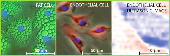
We choose two types of cell — a bovine endothelial cell taken from the aorta and a mouse adipose, i.e. fat, cell. Endothelial cells play an important role in physiology of blood vessels and are useful in the study of biomechanics. Fat cells provide an interesting comparison because of their different geometry.
Fluorescence micrographs of the fat and endothelial cells superimposed on differential-interference- and phase-contrast images, respectively. The nuclei are stained blue in the micrographs. The right-hand image is a picosecond-ultrasonic image of a single endothelial cell with 1 micron lateral and 100 nm depth resolutions. Deep blue corresponds to the lowest ultrasonic amplitude.
The cells are fixed in-vitro to a titanium film on a sapphire substrate, and we use a focused ultrashort-pulse laser to generate and image high-frequency gigahertz ultrasonic pulses travelling through them. A movie of the wave amplitude passing through the cells shows how the ultrasonic waves are more highly damped there.
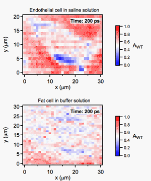
Animation of 10 GHz ultrasonic waves travelling through approximately 0.7 microns of cell tissue. These movies are obtained by wavelet transforms of the raw optical reflectivity change data. Deep blue corresponds to the lowest ultrasonic amplitude.
From these movies we have derived the average physical properties of the cells, such as elastic modulus and ultrasonic attenuation, as a function of depth.
Following this approach for the first 3D imaging of single biological cells with picosecond ultrasonics, it should be possible in future to non-invasively reveal the internal structures of cells in-vivo with 100 nanometer depth resolution. See 'Three-dimensional imaging of biological cells with picosecond ultrasonics,' S. Danworaphong et al., Appl. Phys. Lett. 106, 163701 (2015).